ABSTRACTS
Some abstracts that might be of interest
 |
VISION
RESEARCH ABSTRACTS Some abstracts that might be of interest |
Phaco-Emulsification Causes the Formation of
Cavitation Bubbles
Bengt Svensson
(Umea, Sweden) & John Mellerio
Current Eye Research,
13:649-653, 1994
There have been reports of complications arising
from damage to non-lenticular ocular tissue during the increasingly popular
procedure of cataract extraction with phaco-emulsification. One cause of this
damage might be the formation of cavitation bubbles. Such bubbles are known to
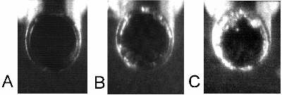
produce free radicals and shock waves. This paper demonstrates directly the
formation of cavitation bubbles at the tip of the phaco-probe. It also shows the
importance of a smooth probe profile in reducing bubble formation.
Recommendations are made for probe and tip design and for the use of minimum
power during the surgical procedure of phaco-emulsification.
The figure shows the end of the phaco-probe: A,
at the end of the advance stroke; C, at the end of the retraction stroke - note
cavitation bubbles. B is half way through the advance stroke. Frequency was
44kHz .
Traffic Signal Light Detection
through
Sunglare Filters of Different Q Factors
David A Palmer, John Mellerio & Amanda
Cutler
Color Research & Application, 22:24-31, 1997
Recent guides for use of sunglare protection filters have introduced the
concept of Q factors as a measure of colour appearance distortion in viewing
traffic signal lights. The adoption of Q factor values was apparently arbitrary
and not firmly based on experimental data. The manner in which changes in Q
factor affect colour perception and detection of signal lights has been measured
and shows that detection thresholds vary with the Q factor in a manner which can
only partly be explained but which is not independent of the colour of the
signal as assumed in the guides.
LED's VIEWED THROUGH COLOURED
LENSES
John Mellerio & David
Palmer
Proc CIE Symp on Standard Methods for Specifying & Measuring
LED Characteristics , CIE publication x013, ISBN 3900734852, Vienna,
1997
The chromaticities and contrasts of a yellow LED simulating a
traffic signal and of an instrument panel indicator (simulated by mixing light
from two LED's) were calculated for viewing through three popular sunglare
protection or driving lenses. Colour naming and quality rating of the lights
confirmed the theoretical changes in chromaticity and contrast.
For example, one driving lens reduced the contrast of the yellow LED traffic
signal to a third of that experienced with naked eye viewing, and halved the
detection distance in a typical viewing situation.
Designers of systems
that employ LED's should ensure that the visual information which they hope to
convey is not degraded to a dangerous extent for those
who wear sunglasses or
driving lenses.
Macular Pigment Measurement with a Novel
Portable Instrument
MELLERIO, J., PALMER, D.A. &
RAYNER, M.J.
European Association for Vision and Eye Research, Palma
de Mallorca, October 1998
PURPOSE
‘ Hammond, Wooten & Snodderly (1997) suggested
that Age Related Macular Degeneration (ARMD) might be correlated with the
Macular Pigment Optical Density (MPOD)
‘ Thus it may be
clinically useful to know a person’s MPOD in order to prognosticate maintenance
of visual function
‘ Macular Pigment (MP), which is yellow,
is a mixture of carotenoids only obtainable from the diet
‘
Knowledge of a patient’s MPOD could be used to advise about diet in order to
improve protection against long-term oxidative light
damage
‘ A practical screening method for determining MPOD
in a clinician’s office is therefore desirable
PRINCIPLES
‘ Heterochromatic Flicker
Photometry (HCFP) is a well established technique for measuring MPOD (Werner
& Wooten, 1979)
‘ A flickering green light, which is
not absorbed by MP, is matched in a test field with an adjustable flickering
blue, which is absorbed by MP (the green and blue lights flicker in counter
phase)
‘ The matches are made either with the retinal image
of the test field on the fovea (Case 1) or on the parafovea (Case 2)
outside the pigmented area
‘ At the match point of minimum
perceived flicker the brightness of the blue and the green will be the
same
‘ The blue light is absorbed by the yellow MP so in
Case 1 the subject needs more blue than in Case 2
‘
Provided that the green is constant it follows that:
MPOD = log B1 - log
B2
where B1 and B2 are the mean blue luminance
values
‘ The flicker frequency must be high enough not to
stimulate the rods - above about 12 Hz
‘ The blue-green
match is made with the medium and long wave cones which are assumed to be evenly
distributed across the central areas of the retina
‘ Any
contribution of the short wave cones and the rods to the matches is prevented by
adapting them with a blue background 
DEVICE
‘ A novel method of obtaining
the stimuli and background is to use Light Emitting Diodes (LED’s) as
sources
‘ The drawing and photographs show a small portable
prototype device using LED’s
‘ The test field, 0.5 deg
diameter, is formed by transilluminating a circular aperture behind which is a
small integrating box containing the blue and green LED’s 
‘ The 5 deg adapting background is formed by an array of
seven blue LED’s filtered to narrow the bandwidth: the light is passed through a
partial diffuser and projected through a field lens before reflection from a
glass plate in front of the test field
‘ The
luminance of the test field is about 50 cd.m-2 and that of the background is
about 10 cd.m-2
‘ The flickering diodes are supplied by a
constant current source developed by Millar and Barnet (QMW College, London):
this source has been carefully designed so that the LED luminances are
approximately linear with respect to the control scale values: Figure 1 shows
the calibration curve for a green LED
‘ Figure 2 shows the
normalised spectral power distributions of the three LED types used in the
prototype together with the absorption of macular
pigment
RESULTS
‘ The measures of
MPOD show distinct differences between individuals which are consistent from day
to day: a few subjects have also been measured by an objective method which
yielded values similar to those from the LED prototype
‘
The mean MPOD for 12 subjects (ages 19 to 65) was 0.287 ±0.136 sd which is
similar to reports in the literature
‘ Figure 3 indicates a
gender difference (p = 0.023) with male MPOD lower than that of females,
contrary to previous reports (Hammond et al, 1996)
‘ Some
subjects were good at making HCFP matches but even poor subjects could obtain
useful results with a minimum of ten pairs of fovea/parafovea observations
CONCLUSION
‘ By using a novel combination of cheap, commercially
available LED’s, a small integrating box and semi-Maxwellian background
projection, we have made a practical system for measuring MPOD which is small
and portable
‘ With appropriate development this would be
suited to automated clinical screening in a wide range of
environments
References
Hammond,
BR, et al. Individual variations in spatial profile of human macular
pigment. J. Opt. Soc. Am. A., 14:1187-1196,
1997
Hammond, BR, et al. Sex differences in macular
pigment optical density, etc. Vision Res, 36:2001-2012,
1996
Werner, JS & Wooten, BR. Opponent chromatic
mechanisms etc. J. Opt. Soc. Am., 69:422-434,
1979
THE DESIGN OF EFFECTIVE OCULAR
PROTECTION FOR CW RADIATION SOURCES
in: Measurement of
Optical Radiation Hazards: A Reference Book International
Commission on Non-Ionising Radiation Protection,
CIE Publication x016-1998,
ISBN 39804789-5-5, 1998
Of the three strategies for ocular protection -
avoidance, reduction of exposure time and attenuation of radiation at the eye -
the last is the most popular for relatively less intense CW sources. It is also
the most difficult to design because not only must it protect safely against
chronic accumulative effects that may take many years to become apparent, it
must allow at least some visual function to be maintained. The physical
realisation of this protection is usually some form of lens fitted to a goggle,
visor or spectacle. The designer must consider the spectral power distribution
(spd) and likely duration of the radiation, and
its irradiance in absolute
units. He must also take into account the action spectrum of each ocular
structure and compare this with the spd to see if the effects of the radiation
are likely to be harmful. If they are, he must then specify a filter with a
spectral transmittance that reduces the transmitted radiation to safe
levels.
It is this process that is a challenge for standards committees
seeking to develop standards criteria for ultraviolet, visible or infrared
protection against solar radiation for use in sunglasses and prescription
eyewear. In this area there is continuing debate on whether any protection is
essential or whether it should be left to the manufacturer to voluntarily build-
in protection. However, with the advent of health criteria and occupational
exposure limits, it has been possible more readily to specify spectral
attenuation factors for welding, cutting, arc-lamps and foundry operations.
There have been debates on the level of specification to spectral detail in such
efforts but the problems seem more tractable.
Whatever the radiation
source for which protection is sought, there follow two further stages in the
specification of a protecting filter. The first is to ensure that the spectral
transmittance of the filter is physically realisable and stable, and secondly to
ensure that the attenuation of the visually effective radiation (light) is not
such as to reduce visual function to below acceptable limits.
In this
paper these stages will be considered in the light of attempts to produce
standards, and attention will be given to the way that safe filters may
prejudice visual performance by reducing contrast thresholds, visual acuity and
color recognition. There is always the danger that the reduction of one hazard
is replaced by another especially if standards are oosely drawn up.
LIGHT-EMITTING DIODES (LEDs) AND LASER
DIODES:
IMPLICATIONS FOR HAZARD ASSESSMENT
International Commission On Non-Ionizing Radiation
Protection (Health Physics,
77:218-220, 1999)
This statement is based upon the
deliberations of the ICNIRP Standing Committee IV ("Optics") and was extensively
discussed in a task group meeting of experts convened by ICNIRP which took place
on 23-25 September 1998 at the University Eye Clinic, Regensburg, Germany. The
following experts participated in this meeting:
W. Cornelius (Australia), D.
Courant (France), P. J. Delfyett (USA), S. Diemer (Germany), W. Horak (Germany),
G. Lidgard (UK), R. Matthes (Germany), J. Mellerio (UK), T. Okuno (Japan), M. B.
Ritter (USA), K. Schulmeister (Austria), D. H. Sliney (USA), B. E. Stuck (USA),
E. Sutter (Germany), and J. Tajnai (USA).
CONCLUSIONS AND RECOMMENDATIONS
It is concluded that all surface
emitting LEDs and IREDs will be judged safe by applying the ICNIRP ELs for
incoherent radiation as well as by the recommendations of CIE TC 6-38 (Lamp
Safety) for realistic viewing conditions. This conclusion applies to any LED
device which does not have optical gain. Only because of the extraordinary
worst-case assumptions built into some current product safety standards, could
one reach the conclusion that an LED or IRED poses a retinal hazard. On the
other hand, the use of laser ELs to evaluate LEDs could result in an
understatement of the lenticular risk if the source is very large and the lens
becomes overheated.
It is therefore recommended that safety evaluations
and related measurement procedures for LEDs follow the guidelines for incoherent
sources (ICNIRP 1997). This approach provides the most accurate assessment of
incoherent sources without problems originating from certain underlying
assumptions incorporated into the limits developed for collimated laser beams.
Diode lasers and VCSELs clearly should be treated in all standards as
lasers.
It is recognized that the determination of appropriate
viewing durations and distances under different conditions of use is needed for
any optical radiation hazard assessment. Unfortunately, not all safety
guidelines currently recommend use of the same measurement distances and viewing
durations. The future development of application-specific safety standards which
may be applied to realistic viewing conditions will also contribute to reducing
unnecessary concerns regarding LED and IRED safety.
International Commission on Non-Ionizing Radiation
Protection (ICNIRP).
Guidelines on limits of exposure for
broad-band incoherent optical radiation (0.38 to 3 :m)
Health Phys. 73:539-597; 1997.
Characteristics of Macular Pigment May be Used to Measure Pigment Density Psychophysically
EXECUTIVE SUMMARY OF THE SPECIAL INTEREST
SYMPOSIUM:
Xanthophyll Carotenoids and the Macular
Pigment
European Association for Vision and Eye Research
Conference
10 – 13 October 2001, Alicante, Spain
Professor Mellerio
explained, “The mixture of lutein and zeaxanthin that is macular pigment has
well determined optical characteristics.” Macular pigment density can easily and
non-invasively be measured in the human eye. Most of the different measuring
techniques available rely on the principle that blue light is absorbed by the
yellow macular pigment i.e. the more yellow pigment present, the more blue light
is absorbed.
Various psychophysical techniques are utilised to measure
the quantity of pigment in the living eye. These include colour matching, motion
anomaloscope, spectral sensitivity and heterochromatic flicker photometry (HCFP)
which is the most common principle employed in these psychophysical methods.
Professor Mellerio presented a portable instrument that can be used to measure
macular pigment densities in field studies or in the optometrist’s or
ophthalmologist’s practice.
MACULAR PIGMENT OPTICAL DENSITY MEASUREMENTS WITH A
PORTABLE SCREENING INSTRUMENT
MELLERIO J1, van
KUIJK E2, PAULEIKOFF D2, AHMADI-LARI S3, MARSHALL J3
1.
University of Westminster, London (UK)
2. Institute of Ophthalmology,
London (UK)
3. Dept. Ophthalmology, St Thomas' Hospital, London
(UK)
(See MELLERIO, J.,
AHMADI-LARI, S., VAN KUIJK, F.J.G.M., PAULEIKHOFF, D., BIRD, A.C. &
MARSHALL, J. A portable instrument for measuring macular pigment with central
fixation. Current Eye Res, 25:37-47, 2002 for
full publication of this work)
European Association for Vision and Eye
Research Conference
10 – 13 October 2001, Alicante, Spain
Purpose To see if macular pigment (MPOD) in the eyes of normal
subjects and AMD patients varies with age, gender, eye colour, smoking habit,
diet, chronic exposure to sunlight and disease state of the
retina.
Methods The portable screening instrument (described at
EVER 1999) uses light emitting diodes and a free viewing condition, and employs
heterochromatic flicker photometry. 107 subjects with normal vision
measured MPOD in each eye and answered a simple questionnaire designed to find
their age, gender, eye colour, smoking habit, what diet they ate and their
exposure to sunlight. These last two parameters were only crudely reported
as more or less than 16 servings of vegetables, fruit and eggs per week and less
than 3 hours on average per day exposure to sunlight or more than 3 hours plus a
sunbathing habit. MPOD was also measured in 17 patients with AMD and in
age and gender matched controls.
Results The mean MPOD for
healthy subjects was 0.41±0.16 sd. There was no significant correlation of
MPOD with age but there were significant differences (p<0.05) for gender,
iris colour and smoking habit similar to those reported in the literature.
The group of subjects with the diet poorer in vegetables, fruit and eggs had a
significantly smaller MPOD than those with a richer diet. Subjects with a
greater exposure to sunlight had significantly less MPOD than those with the
smaller exposure. Male patients with advanced AMD had significantly less
MPOD compared to those with healthy eyes and although the female eyes showed a
similar trend, the difference did not reach
significance.
Conclusions The findings support those reported in
the literature for age, gender, eye colour, smoking habit and richness of diet
in carotenoids. The reduction of MPOD with sunlight exposure and with
advanced AMD in males is interesting and requires further
investigation.
| Return to the VISION RESEARCH | 
|
End of Abstract Page
Any comments and suggestions for
improvements
are welcome to the page editor via email at: